Necropsy methods
Information from carcasses which may be useful
for loris
and potto conservation
Still under construction -
incomplete
first draft. Update here soon. Any help for correction would
be appreciated
Compiler: Helga Schulze; with contributions by (in alphabetical
order):
Eberhard Curio; Colin Groves; Anna Nekaris; Kathrin Petry;
Heinz-Adolf
Schoon; Roland Plesker; Christian Roos; Ulrike Streicher. The
initial draft
was based on some publications, for instance Wobeser and Spraker
1980:
Post-mortem examination, Munson 2000: Necropsy procedures for wild
animals,
and others (see references).
This review is partly based on general mammal necropsy
guidelines
which probably rather consider procedures for larger species. So
some of
the described procedures may not be adequate for small species
like slender
lorises, but as far as possible, methods are adapted. If not
only a veterinarian
examination for diseases and causes of death is planned, but
also preservation
of specimens for anatomical or taxonomic collections, a choice
between
identification of health problems and other aims will be
necessary because
veterinarian examination is usually connected with destruction
of structures.
This page may be regarded as a list of propositions to chose
from for certain
purposes.
Introduction: information
which
can be obtained from carcasses of animals and other material found
in the
wild may be very useful for obtaining data for conservation.
Therefore,
some considerations before starting a necropsy will be useful. In
addition,
the form of necropsy reports and
their
possible role as official documents may be important
in law suits
and should be considered by an appropriate form of records and
resulting
documents. Therefore preparation of a form sheet prior to necropsy
is recommended.
See also: information about recording
of
data,labelling,
preservation and storage of carcasses and samples.
Necropsy should take place as soon as possible. If no quick
examination
is possible, smaller carcasses, after noting all necessary data,
should
be kept cool, but nor frozen (AZA Prosimian Advisory Group, 2002),
if possible.
See also below: dealing with autolytic or
decomposed
carcasses.
A good necropsy procedure is supposed to allow identification and
examination
of each organ, including opening of all hollow organs. The less
experience
a prosector has, the more rigidly he / she should adhere to a
standard
technique to assure that all organs are examined thoroughly.
Becoming familiar
with the normal appearance of organs will then help to easily
recognize
abnormalities.
For evaluation of the findings, a cooperation with biologists,
specialized
pathologists and diagnostic laboratories or experts from
Universities or
other institutions may be necessary (Wobeser, Spraker 1980).
Equipment: see extra file (in
preparation)
First records, sample collection and
measures
in the place where an animal has been found (in
preparation)
Self-protection before and during examination:
Protective clothing: (in preparation)
Formalin is highly toxic; it should be handles with great care,
and
hands should be washed thoroughly when work is finished
(Rabinowitz et
al., 2000).
Procedures in cases of dangerous infectious diseases (in
preparation)
Protective clothing and method
for
examination of brains in cases of rabies: see below
Samples from autolytic or decomposed carcasses
Autolysis of tissues (= self-dissolution after death due to action
of own enzymes) can cause many artifacts in tissues that can
superficially
be confused with a disease process, but histopathological
examination can
still distinguish between true lesions if tissues are properly
handled.
For histopathology, autolyzed or decomposed tissue samples must be
handled
very gently, held at the edges only, cut with a sharp knife or
scapel and
quickly placed in formalin (freezing would further diminish the
ability
to recognise most changes microscopically). Samples both from
changed-looking
and more normal appearing parts of an organ will be most useful.
Quick
cooling or preservation in formalin is recommended (one of us: K.
Petry).
For infectious disease or toxicology testing, freezing or
refrigeration
of samples should take place soon as possible (Munson, 2000).
Examination of dead neonates:
In addition to adult necropsy as described below, some additional
examinations
are recommended:
Has breathing occurred? (do the lungs float in formalin?) (Munson
2000)
Umbilical stump - sample including surrounding tissues (Munson 2000)
Signs of dehydration / tissue moistness?
Milk remnants in the gastrointestinal tract?
Malformations such as cleft palate, deformed limbs?
Evidence that the baby has fallen down (found on the ground,
lesions,
broken limbs)?
(Examinations of embryos:
see below)
Necropsy
External examination of the animal (based
on Munson
2002; one of us: K. Petry; one of us: H.-A. Schoon; one of us: R.
Plesker)
External parasites? (Preservation of some)
Posture, description / photos
Rigor mortis, other signs of death, externally visible signs of
decomposition?
Blood visible? If so: clotted, dried?
Wounds? If so: signs of bruising or bleeding into the tissue,
indicating
that the wounds occurred when the animal was still alive?
(Otherwise they
might be signs of scavenging). Cause of wounds?
Externally visible: parts of the body missing, changed? Broken
bones,
missing hair, broken teeth, other signs of trauma?
Hair, skin, nails: changes, missing?
Malformations?
Weight, head
and body
measurements
Nutritional state: externally visible fat
stores, muscle mass? Dehydration?
Sex, external
signs
of reproductive status:
Males: testes
scrotal
or inguinal, testes size, scrotal skin patterns, other
observation
(secretion)
Females: Conditon of the vagina: open,
closed, sealed by a skin? A sealed vaginal cleft may occur
in juvenile
females and in seasonal breeders at certain times of the year,
with the
vaginal opening closed by a membrane which may look like normal
skin with
vaginal opening absent (Nagorsen, Peterson, 1980; observation at
Ruhr-University).
Signs of estrus such as swollen and reddened rims of vaginal
opening?.
Signs of pregnancy? Mammary glands: signs of lactation? (Can milk
be squeezed
from the teats?) Nipples looking used? (Nagorsen, Peterson 1980)
Pregnancy? In Loris, pregnancy is usually inconspicuous,
even
highly pregnant females may show a dilated ribcage rather than an
enlarged
belly (captive observation at Ruhr-University).
Offspring: babies / infants clinging to the adult found, babies /
infants
found parked in the proximity?
Condition of the teeth
External examination of eyes, ears before opening the carcass.
Blood,
pus, parasites in the ears? Blood in the ear and haemorrhage in
the skin
around the eyes may be signs of a skull base fracture.
Hair samples
may be taken for DNA analysis or for a taxonomical reference
collection
Samples for microbial examination from externally visible
abnormal areas
(for instance pus, sample from areas with abscesses, samples from
the edge
of affected (abnormal) areas most likely to contain organisms)
should be
taken with sterile instruments and stored in sterile
container (Munson
2000)
Planning what to preserve, prior to dissection
After external examination, a decision is necessary whether
preservation
of the entire specimen, preservation of parts for non-veterinarian
purposes
or dissection is most important. This decision must be dependant
on importance
of destruction of parts for examination (for instance if diagnosis
is important
because of danger of epidemics or zoonoses) or importance of
preservation
of parts of the specimen (skull, skin, other) or the whole
specimen, for
instance for taxonomic purposes.
Dissection
Primates (all species) and small mammals are dissected while lying
on the back; they may be fixed in this position. Special devices
for laboratory
rodents, a soft board, sheet of cardboard or styrofoam covered
with plastic
foil and four stout pins or hypodermic needles; (Wobeser and
Spraker, 1980;
Schoon, lecture manuscript).
1) Skin Preservation of the skin necessary for mounting or preparation
of
a study skin planned? If so, the inner skin surface with
hair roots
should not be damaged when removing the skin from the body;
otherwise the
mounted specimen may later loose hairs. In carcasses beginning to
decompose,
the hair may already be loose (“slipping” fur), it will then fall
out leaving
a naked skin unless removal of the skin is done very cautiously.
Therefore,
tests (trying to plug a few hairs in several parts of the body
including
the abdomen) prior to skin removal should be done in taxonomically
valuable
specimens.
Removal of skin for study skins or mounted specimens (in
preparation), preparation of
a study
skin (in preparation),
mounting a skin (in preparation).
Removal of skin prior to necropsy may also be useful for other
reasons.
If
necropsy is not done immediately, removal of the skin with
insulating fur
before cooling or freezing may help to cool the carcass down more
quickly.
In cases with superficial tumours or lesions supposed to be
examined, it
may be necessary to leave the parts of skin concerned attached to
the body
(Schoon, lecture manuscript).
For dissection, the ventral side is opened with an initial
skin
incision with sterile instruments, usually from the lips along the
ventral
midline to the perineum, passing the penis and scrotum or clitoris
on one
side. If the skin is supposed to be preserved as a study skin or
mounted
specimen, it will be useful not to cut trough too much hair
(moistening
of the hair may help to prevent it from moving between the
scissors); it
may be useful to avoid cuts in thinly haired parts of the skin,
where after
mounting ugly seams might be visible, rather leaving parts of the
limbs
in the skin. (Vacuum freeze-drying with limbs in the skin may be
an adequate
method for mounting thinly-haired specimens like slender lorises).
An examination of the inner surface of the removed skin may show
wounds
hidden under the hair or effusions of blood. In addition, muscles,
fat
and mammary glands can be examined on this occasion. Mammary
tissue is
usually whitish in colour, beneath the teats under the skin;
presence of
milk should be noted (Nagorsen, Peterson 1980). The glands and
draining
superficial lymph nodes can be incised for examination of the
parenchyma
(Wobeser, Spraker, 1980).
2) Opening of the body cavities: general
All body cavities (abdomern, chest, heart)
must
be opened. Technique see below.
Sterile samples of organs for microbial
examination
should be taken for culture with sterile instruments and stored
in sterile
containers before organs are handled (Munson, 2000). Such
samples will
be needed for instance from all abnormal-looking areas, for
instance from
pus, from areas with abscesses or nodules, from the edge of
affected (abnormal)
areas most likely to contain organisms.
Small amounts of blood can be
collected
on filter paper and used in testing for some viral, bacterial
and blood
parasite DNA or RNA (blood
sampling methods:
in preparation). Antibodies against
some pathogens
may also be detected using these samples.
Initial notes. In the opened body cavities, before any
organs
are removed, the following should be noted:
General condition
Smell
General nutritional condition of the animal, fat deposits
Signs of dehydration?
Condition of the serous membranes (membranes covering the inner
surfaces
of body cavities), pleural membranes? (Wobeser, Spraker 1980;
Schoon, lecture
manuscript)
Location of all organs: any organs displaced? Abnormal attachment
of
any organs to the body cavity? (Munson, 2000).
Abnormal fluids in the pleural, pericardial, abdominal cavity? If
so:
quantity, colour, smell? Collection of liquid samples.
(Examination later
in the lab: ascites, tumor cells? What do the granulocytes (white
blood
cells) look like: juvenile or with polymorphic nuclei? One of us:
K. Petry)
See also below: examination
of
the body cavity after removal of organs
Sections of all lesions and unnormal-looking parts should be
collected
during following dissection (Munson, 2000).
Opening of the abdomen
The abdominal wall is opened by cutting along the ventral midline
from
pelvis to sternum. After two additional cuts along the lower rim
of ribcage
the resulting flaps can be deflected (Wobeser, Spraker 1980;
Schoon, lecture
manuscript).
Opening of the thorax
If no skeletal preservation for a collection is necessary, the
thorax
may be opened by cutting through the ribs along both sides (in
small mammals
with bone scissors, for larger species a saw would be necessary)
and removal
of the sternum with attached parts of ribcage (Wobeser, Spraker
1980; one
of us: R. Plesker; for large mammals, the procedure would be
different,
see Wobeser, Spraker, 1980 or Munson 2000). Alternative: a cut
along the
region where the ends of rib bones are connected to cartilage,
where cutting
is easier and the bony parts of skeleton are less damaged (one of
us: K.
Petry).
Use of the removed ribs for examination of the stability
(ossification)
of bones and bone marrow examination: see below, under "examination
of muscles and skeleton".
Opening and examination of the oral cavity and throat
Cuts along the inner margins of the mandible are necessary to free
the tongue. Then the larynx, hyoid bone and other structures can
be freed
by cautious traction in cranial direction and cutting down to the
soft
gums (Palatum molle); the throat organs are freed caudally up to
the cranial
thoracic opening. Muscles and ligaments at the mandibular joint
should
be cut through; disarticulation of the mandible and cautious
cutting free
of the cheek musles and mucosa from the lateral surfaces of the
mandibular
bone then allows a complete examination of the oral mucosa
(Wobeser, Spraker
1980; Schoon, lecture manuscript).
The inside of the mouth, tonsils, and teeth should be examined.
Erosions,
ulcerations or other lesions on the oral / pharyngeal mucosa,
tonsils or
any other areas in the mouth? The salivary gland should be
examined; the
tonsils and several lymph nodes under the skin (at the angle of
the jaw
and above the larynx) should be removed for histology (Munson,
2000)
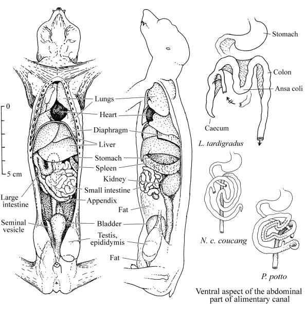
Figure: left: ventral and lateral aspect of the inner organs of
a male
slender loris (probably L. t. tardigradus or an
intermediate L.
t. tardigradus / grandis form).
Right: half-schematic ventral aspect of the abdominal part of
alimentary
canal in Loris, Nycticebus and Perodicticus
(Arctocebus:
similar to Loris), based on Osman Hill and Rewell 1948,
Osman Hill
1953 and 1972.
Superficial examination of the thoracic viscera
A sample of lung tissue for bacteriology should be taken (Schoon,
lecture
manuscript)
Examination of tongue, tonsils and the pharyngeal area (Wobeser,
Spraker
1980).
External surfaces of the lungs: any lobular differences in the
lungs
such as different colour, firmness or inflation should be noted
(Wobeser,
Spraker 1980). Edema, emphysema? (Munson 2000).
Presence and size of thymus? (Wobeser, Spraker 1980)
Checklist: recommendations which fixed
tissue
samples for histology + other samples should be collected /
preserved:
in preparation
In carcasses of rare species, preservation of organs in situ
for anatomical studies may be useful, with only small samples
taken out
for examination, causing as little disturbance as possible. If a
necropsy
is only done for detection of health problems, enough tissue for
examinations
including bacteriology should be taken (one of us: K. Petry),
samples including
abnormal areas and surrounding normal areas; Munson (2002)
recommends samples
no thicker than 1 cm (for good fixation), but long and wide enough
to represent
different areas of a tissue and possible abnormalities. In small
animals,
entire organs instead of samples may be collected.
Heart
Before removal of organs, the heart can be opened with sterile
tools
to take a blood sample for culture (the right atrium is the best
location),
then additional blood can be taken to obtain serum for serological
tests.
Is fluid found in the pericard after opening it? The heart and
great
vessels should be examined while still attached to the lungs (for
easy
reorientation in case of some anomalous condition of the vessels).
The
epicardial surface, size and contour of the heart should be
inspected (Wobeser,
Spraker 1980). Schoon (lecture manuscript) recommends to take the
following
measurements: circumference of the heart in the atrioventricular
groove
(Sulcus coronarius), height of left and right ventricle.
Opening of the heart: usually by following the normal path
of
blood flow, beginning in the right atrium. A u-shaped incision is
made
through the right atrio-ventricular valve, following the
inter-ventricular
septum to the apex, then back to the base of the heart through
pulmonary
valve. The ventricular wall flap, cut loose this way, can be
lifted to
expose the ventricle and valves. The left side of the heart can be
opened
in a similar way, the incision beginning in the left atrium,
passing through
the atrio-ventricular valve to the apex and back through the
aortic valve
into the aorta (Wobeser, Spraker 1980; one of us: K. Petry,
Schoon, lecture
manuscript).
In recently dead animals, the right heart usually contains plasma
clots
(yellow / tan material), unclotted blood, or clotted blood
(Munson, 2000).
Postmortal blood clots inside the heart should look smooth;
rough-looking
blood may indicate a thrombosis (one of us: R. Plesker). See
collection of blood or plasma. A yellowish inner surface of
the aorta
is a sign of jaundice (one of us:: R. Plesker).
Removal of the heart: see below:
Examination, removal of organs. Systematic
collection
of samples
Removal, examination of the heart
If no changes in the blood vessels have been noticed during
opening
and first examination, the major bloodvessels can then be cut for
removal
of the heart, close to the lungs, the aorta and posterior vena
cava close
to the diaphragm, remaining connected with the heart. Examination
of the
heart should include thickness of walls (right, left ventricle,
septum),
the inner surface (papillary muscles) the entire septum muscle
(cut), and
collection of samples: several sections of ventricle muscle,
particularly
close to the papillary muscles and rhythmic center (Schoon,
lecture manuscript)
Removal of the cranial parts of digestive and respiratory
tract and
other organs from throat and thorax
The ends of the esophagus should be closed with two adjacent
ligatures
(= threads tied around it) and then cut between adjacent ligatures
to prevent
leakage of contents into the body cavities (one of us: K. Petry;
Schoon,
lecture manuscript; Wobeser, Spraker 1980). It can then be
transected at
the diaphragma; then tongue, larynx, thyroids, parathyroids,
trachea, esophagus,
lungs and thymus can be removed (Wobeser, Spraker 1980);
Examination of these parts after removal
The tongue should be examined, and a cross section near tip
including
both mucosal surfaces should be collected (Munson, 2000). If
skeletal preparation
is planned, preservation of the bony parts of larynx (hyoid bone)
must
be considered (Piechocki 1986). For examination, larynx and
trachea should
be opened by a dorsal longitudinal incision with scissors
continueing to
the tips of the diaphragmatic portion along the major bronchi and
into
some smaller bronchi (Wobeser, Spraker 1980; Schoon, lecture
manuscript).
Exsudate in the airways? (Wobeser, Spraker 1980). Parasites such
as trematodes?
(Nagorsen, Peterson 1980). Transverse sections from several lobes,
including
a major bronchus and trachea, should be preserved (Munson, 2000).
The bronchial
lymph nodes should be examined after an incision (Wobeser, Spraker
1980).
Changes in the lungs such as emphysema are not necessarily a sign
of
disease, they can also be a consequence of death or euthanasia
(one of
us: R. Plesker).
The following glands and lymph nodes should be examined:
Thyroid / parathyroids (Munson, 2000)
Lymph nodes (cervical, mediastinal, bronchial, mesenteric and
lumbar),
cut transversely (Munson, 2000).
Thymus (present only in young animals) (Munson, 2000; one of us:
K.
Petry)
The diaphragm contains muscle cells, a sample may be
collected
for muscle examination, see under "muscles"
(in preparation)
Liver and gall bladder
The liver may be removed from the body cavity together with the
first
part of duodenum, cutting the ligament and blood vessel
connections to
the diaphragm without damaging the latter. Examination whether the
bile
duct is open is possible by slightly pressing on the gall bladder
after
opening the duodenum (Schoon, lecture manuscript). This is
particularly
important if the liver looks yellowish (one of us: R. Plesker).
Tha gall
bladder should be examined for stones (one of us: R. Plesker). If
examination
for a Salmonella infection is necessary, the gall bladder
should
not be opened during dissection (Schoon, lecture manuscript).
Trematode
parasites may occur in the liver (Nagorsen, Peterson 1980). After
lasting
environmental distress in captivity, in lorises death due to fatty
liver
and liver necroses has regularly occurred (data from institutions
cooperating
with Ruhr-University).
Sections from 3 different areas of the liver including gall
bladder
should be collected (Munson, 2000); storage of some liver tissue
samples
for DNA analysis (few g, in plastic bags, frozen) for genetic
evaluation
are recommended (AZA Prosimian Taxon Advisory Group, 2002).
Spleen: a cross sections including the capsule should be
preserved
and examined (Schoon, lecture manuscript; Munson, 2000).
An enlarged spleen may indicate a longerlasting infection,
duration
maybe a week or longer (one of us: R. Plesker)
Gastrointestinal tract
Before removal of the intestine from the abdominal cavity, liver
and
gall bladder with bile duct leading into the intestine should be
examined,
and closing of its sections with two adjacent ligatures (= threads
tied
around it) on either side is recommended, then it can be cut
between the
ligatures. Places to be closed this way: both ends of esophagus
(caudally
close to the kardia = entrance of stomach), both ends of the
stomach (one
of us: K. Petry), duodenum cranial from the ligamentum
duodenocolicum =
connection between duodenum and colon and anal end = rectum (one
of us:
K. Petry; Wobeser, Spraker 1980; Schoon, lecture manuscript).
Before removal
of the intestine, liver and gall bladder with bile duct leading
into the
intestine should be examined. For removal, the esophagus can be
transected
at the diaphragma, and tongue and esophagus, lungs and thymus can
be removed
together with other organs in the head and throat region such as
respiratory
tract and glands (Wobeser, Spraker 1980). The intestine can be
separated
from the mesenthery, taken out and spread for examination (Schoon,
lecture
manuscript), or liver, spleen and digestive system can be taken
out as
a block. Stomach and intestine remain closed at both ends and are
opened
last (Wobeser, Spraker 1980).
If a carcass is found in the wild, collection of the content
of
the entire alimentary tract for food analysis may be useful.
Examination
of the stomach content alone may not be sufficient for this
purpose; in
galagos and pottos, after gum-eating it seems that gums are
retained in
the stomach only for few minutes, so usually no trace of gum is
found there,
but gum may be found in the caecum (Hladik 1979, partly quoting
Charles-Dominique
1971 and 1974). Contents of the digestive tract can be preserved
in 5%
formalin or 30-40% alcohol (Rabinowitz et al., 2000), or the whole
alimentary
tract may be preserved in formalin, after injection of formalin
into the
stomach, for later analysis (one of us: A. Nekaris).
Pancreas: before separating the pancreas from the
intestine,
the duct leading from the pancres into the duodenum may be
examined. Munson
(2000) recommends to preserve cross sections from two areas of the
pancreas
together with the duodenum. This may be a method for larger
species. Preservation
the of entire pancreas may be more adequate. In this case, curling
or shrinking
of the soft pancreas tissue can be prevented by spreading the
whole organ
on tissue paper after removal, cautiously folding the paper around
it and
depositing the whole package in fixative; the paper will keep the
tissue
spread out in normal shape during fixation (one of us: R.
Plesker).
Examination of intestine:
The examiner must descide whether the entire content of the
intestinal
tract should be preserved for food analysis or whether other
examinations
may be more important. Before preserving the intestine or
longitudinally
opening it for examination, sections from different areas may be
closed
with thread and removed for later laboratory examination (Schoon,
lecture
manuscript).
Preservation of samples from the intestine should include
multiple
sections from different areas, about 3 cm2 (Munson,
2000) of
the following parts:
Duodenum (part of intestine following the stomach): White and
Edwards recommend
to take two sections of the duodenum together with pancreas (in
larger
animals?); in smaller species like lorises or pottos,
preservation of the
entire pancreas may make more sense; see below, under pancreas.
Jejunum
Ileum (section close to caecum)
Caecum
A piece of rectum with content for test for parasites (one of
us: R. Plesker)
and pancreatic insufficiency (one of us: K. Petry)
Omentum (Munson, 2000)
Intestinal lymph nodes from the middle of the mesentherium (one of
us: R. Plesker)
Stomach: for examination, longitudinal opening along the
Curvatura
major = greater curvature is recommended (one of us: R. Plesker;
Schoon,
lecture manuscript). The gastric mucosa is examined for ulcera,
erosions
and parasites? (One of us: K. Petry). After lasting environmental
distress
in captivity, in lorises bleeding ulcera in the stomach and
intestine have
been found (data from institutions cooperating with
Ruhr-University).
Urogenital system
After removal of the gastrointestinal tract and liver, the
kidneys,
adrenals, reproductive organs and rectum / anus can be taken out
together,
starting with a cut around the kidneys, from cranial via lateral
to caudal;
the whole system should be cautiously cut loose together with
lymph nodes
and without damaging the urinary passages. For the caudal part,
the ventral
part of the pelvis may be removed with bone scissors if no
skeletal preparation
is planned, starting from the foramina obturatoria. The whole
connected
system can then be taken out and spread on a flat surface for
examination
(Schoon, lecture manuscript).
Enlarged adrenals indicate longer-lasting stress prior to
death
(one of us: R. Plesker). In primates the adrenals in general are
larger
than in most other mammals; veterinarians not familiar with this
may erroneously
believe that normal adrenals are enlarged (one of us: R. Plesker).
The
entire gland should be collected with transverse incision (Munson,
2000).
Kidneys - A longer piece of urinary tract left attached to
the
left kidney during removal can help to distinguish it from the
right one
after separating both from the rest (Schoon, lecture manuscript).
Samples:
cortex and medulla from each kidney. Munson, 2000, recommends two
transverse
cuts (for large species?); one of us, R. Plesker, cuts the whole
kidney
logitudinally for examination.
Urinary passages free? (Can be tested with a probe). Urine sample:
gravel or crystals present? (One of us: K. Petry)
Urinary bladder, ureters, urethra - cross section of bladder and 2
cm sections of ureter and urethra should be collected (Munson,
2000).
Reproductive organs:
Reproductive tract, male: location
of testes (scrotal, inguinal?). Measurements of length and
width of
both testes for assessment of reproductive status (Nagorsen,
Peterson,
1980). Samples from both testes (transversely cut) with epididymis
(Munson,
2000); the size of the tubules in the cauda epididymis and
presence of
sperm can provide information about the reproductive status.
Tubules in
breeding males may be swollen and visible to the eye, if so, this
should
be noted (Nagorsen, Peterson, 1980). Entire prostate, transversely
cut
(Munson, 2000). For taxonomic examination, in males preservation
of the
external genitalia for examination of penis shape, length and
degree of
spininess (Bearder et al., 1996) may be useful. The shape of the
baculum
(os penis) might also be meaningful (Source?)
Reproductive tract, female: sample: entire uterus and
ovaries
with longitudinal cuts into lumens of uterine horns (Munson,
2000).
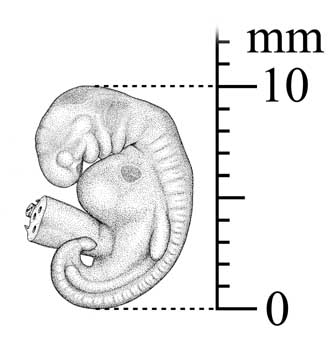
Embryo size measurement (crown-rump length),
taken in natural bent posture (redrawn, changed,
from Nagorsen, Peterson 1980)
.....
Presence, size of embryos? (size:
crown-rump length,
measured like sitting height, but in natural bent
posture).
Possibly resorbed embryos would be conspicuously smaller
and underdeveloped
when compared with normal ones; if so, the condition
should be noted. Embryos
may be preserved in formalin or Bouin´s
solution. In some mammals (for instance in shrews,
rodents and carnivores)
placental
scars remain in the uterine wall after earlier
births, allowing some
conclusions concerning reproductive history of the animal;
such scars are
initially yellow to black pigmented spots on the inside of
the uterus,
later becoming paler and smaller; in mice they may persist
to one year.
For simian primates, Benirschke (2002) provides photos and
describes them
as hyalinized or occasionally calcified scars or plaques
persisting for
months to more than a year. Both the number of detectable
scars and number
of sets of scars of different age should then be noted.
The examined female
can then possibly be classified as nulliparous (signs of
breeding success),
primiparous (with embryos or one set of placental scars)
or multiparous
(with embryos and at least one set of placental scars or
with more than
one set of placental scars) (Nagorsen, Peterson 1980; more
information
possibly from Hackländer et al., 2001).
The reproductive status of partly decomposed animals
should not be guessed.
(Nagorsen, Peterson 1980)
Still incomplete. More included soon
(organ review - eyes ff)
Examination of
the
body cavity after removal of organs:
Inner side of walls / serosa: reddened?
(Sign
of inflammation). Deposits (Fibrin, other)? (One of us: R.
Plesker)
After necropsy, disinfecting the necropsy site is
necessary.
The carcass, all remaining tissues and blood soaked dirt or waste
should
be buried or incinerated. All contaminated paper or plastic
materials should
be either thoroughly disinfected or incinerated. All blood and
residual
tissues should be removed from the instruments and tools with soap
and
water. Then the instruments should be disinfected. Necropsy boots
and apron
should be cleaned and any contaminated clothing thoroughly washed.
The
external surfaces of any containers with samples should also be
washed.
(Munson, 2000)